MPO
HPA
RESOURCES
  • TISSUE
  • BRAIN
  • SINGLE CELL
  • IMMUNE CELL
  • SUBCELLULAR
  • CANCER
  • BLOOD
  • CELL LINE
  • STRUCTURE
  • INTERACTION
  • METABOLIC
  • ISOFORM
  • EVOLUTIONARY
ABOUT
  • INTRODUCTION
  • HISTORY
  • ORGANIZATION
  • PUBLICATIONS
  • ACKNOWLEDGMENTS
  • CONTACT
NEWS
  • NEWS ARTICLES
  • PRESS ROOM
LEARN
  • DICTIONARY
  • PROTEIN CLASSES
  • PROTEIN EVIDENCE
  • METHODS
  • AI IMPLEMENTATIONS
  • EDUCATIONAL VIDEOS
DATA
  • DOWNLOADABLE DATA
  • PUBLICATION DATA
  • RELEASE HISTORY
HELP
  • HELP & FAQ
  • ANTIBODY AVAILABILITY
  • DISCLAIMER
  • PRIVACY STATEMENT
  • LICENCE & CITATION
Fields »
Search result

Field
Term
Gene name
Class
Subclass
Category
Keyword
Chromosome
External id
Tissue
Cell type
Expression
Antibody panel
Tissue
Main location
Patient ID
Annotation
Tissue
Category
Tau score
Tissue
Category
Tau score
Tissue
Category
Tau score
Cluster
Reliability
Region
Niche
Category
Brain region
Category
Tau score
Brain domain
Category
Tau score
Cell type
Category
Tau score
Cell type
Category
Tau score
Brain region
Category
Tau score
Brain region
Category
Tau score
Brain region
Category
Tau score
Brain region
Category
Tau score
Cluster
Reliability
Cell type
Category
Tau score
Cell type group
Category
Tau score
Single cell
Category
Tau score
Single cell
Category
Tau score
Annotation
Annotation
Annotation
Tissue
Cell type
Enrichment
Cluster
Cell type
Category
Tau score
Cell lineage
Category
Tau score
Cluster
Location
Searches
Location
Cell line
Class
Type
Phase
Reliability
Cancer
Prognosis
Cancer
Regulation
Cancer
Regulation
Cancer
Category
Cancer
Category
Tau score
Correlation
Correlation
Cancer
Category
Tau score
Cluster
Variants
Source
PTM
ipTM
Interacting gene (ensg_id)
Type
Number of interactions
Type
Heterodimer
Subsystem
Isoforms
Specificity
Expression
Species
Ortholog
Type
Species
Tissue
Category
Species
Tau score
Data
Category
Data
Species
Category
Category
Category
Category
Category
Validation
Validation
Validation
Validation
Antibodies
Data type
Column


BETA
  • SUMMARY

  • TISSUE

  • BRAIN

  • SINGLE CELL

  • IMMUNE

  • SUBCELL

  • CANCER

  • BLOOD

  • CELL LINE

  • STRUCTURE

  • INTERACTION

  • METABOLIC

  • ISOFORM

  • EVOLUTION

  • MPO
PROTEIN SUMMARY GENE INFORMATION RNA DATA ANTIBODY DATA
Hippocampal formation Amygdala Basal ganglia Midbrain Spinal cord Cerebral cortex Cerebellum Hypothalamus Choroid plexus Retina Thyroid gland Parathyroid gland Adrenal gland Pituitary gland Lung Salivary gland Esophagus Tongue Stomach Duodenum Rectum Small intestine Colon Liver Gallbladder Pancreas Kidney Urinary bladder Testis Epididymis Prostate Seminal vesicle Vagina Breast Cervix Endometrium Fallopian tube Ovary Placenta Blood vessel Heart muscle Skeletal muscle Smooth muscle Adipose tissue Skin Bone marrow Tonsil Appendix Lymph node Spleen Thymus
MPO INFORMATION
Proteini

Full gene name according to HGNC.

Myeloperoxidase
Gene namei

Official gene symbol, which is typically a short form of the gene name, according to HGNC.

MPO
Protein classi

Assigned HPA protein class(es) for the encoded protein(s).

Cancer-related genes
Candidate cardiovascular disease genes
Disease related genes
Enzymes
Human disease related genes
Metabolic proteins
Plasma proteins
Potential drug targets
Protein evidence Evidence at protein level (all genes)
Number of transcriptsi

Number of protein-coding transcripts from the gene as defined by Ensembl.

2
Protein interactions Interacting with 3 proteins according to aggregated data from 1 databases
Metabolic subsystems Involved in 10 reactions in 3 subsystems
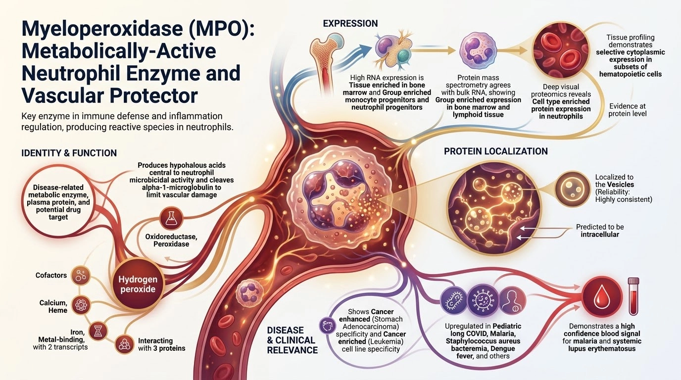
MPO AI GENERATED INFOGRAPHICi

This infographic was generated by AI (Gemini) using only data presented in the Human Protein Atlas v25. It is a quick overview and may contain errors or simplifications, so please check the full data on this gene page before drawing conclusions.

Read more
AI generated infographic for MPO

Disclaimer: This infographic was generated with the help of artificial intelligence, based on data and literature for MPO. It is intended as a simplified visual overview only and may contain errors, omissions or oversimplifications. It has not been individually reviewed by experts and should not be used as a primary source. Please refer to the data presented in the Human Protein Atlas and to the primary literature.

Report an error in this infographic

What seems to be wrong? (select all that apply)
PROTEIN EXPRESSION AND LOCALIZATION
Tissue profilei

A summary of the overall protein expression profile across the analyzed normal tissues based on knowledge-based annotation, presented in the Tissue resource.

"Estimation of protein expression could not be performed. View primary data." is shown for genes where available RNA-seq and gene/protein characterization data in combination with immunohistochemistry data has been evaluated as not sufficient to yield a reliable estimation of the protein expression profile.

Selective cytoplasmic expression in subsets of hematopoietic cells.
Tissue specificity (MS) Group enriched (Bone marrow, Lymphoid tissue)
Cell type specificity (DVP) Cell type enriched (Neutrophils)
Subcellular locationi

Main subcellular location based on data generated in the subcellular section of the Human Protein Atlas.

Localized to the Vesicles In addition localized to the Nucleoplasm
Predicted locationi

All transcripts of all genes have been analyzed regarding the location(s) of corresponding protein based on prediction methods for signal peptides and transmembrane regions.

  • Genes with at least one transcript predicted to encode a secreted protein, according to prediction methods or to UniProt location data, have been further annotated and classified with the aim to determine if the corresponding protein(s) are secreted or actually retained in intracellular locations or membrane-attached.
  • Remaining genes, with no transcript predicted to encode a secreted protein, will be assigned the prediction-based location(s).

The annotated location overrules the predicted location, so that a gene encoding a predicted secreted protein that has been annotated as intracellular will have intracellular as the final location.

Secreted, Intracellular (different isoforms)
Extracellular locationi

All genes with at least one isoform expected to be secreted to the extracellular environment have been annotated and classified either as secreted to blood or as locally secreted, depending on the predicted final location of the corresponding protein. Proteins expected to be locally secreted have been further classified according to their site of expression.

Secreted to blood
TISSUE RNA EXPRESSION
Tissue specificityi

The RNA specificity category is based on normalized mRNA expression levels in the consensus dataset, calculated from the RNA expression levels in samples from HPA and GTEX. The categories include: tissue enriched, group enriched, tissue enhanced, low tissue specificity and not detected.

Tissue enriched (Bone marrow)
Tissue expression clusteri

The RNA data was used to cluster genes according to their expression across tissues. Clusters contain genes that have similar expression patterns, and each cluster has been manually annotated to describe common features in terms of function and specificity.

Bone marrow - Innate immune response (mainly)
Brain specificityi

The regional specificity category is based on mRNA expression levels in the analysed brain samples, grouped into 13 main brain regions and calculated for the three different species. All brain expression profiles are based on data from HPA. The specificity categories include: regionally enriched, group enriched, regionally enhanced, low regional specificity and not detected. The classification rules are the same used for the tissue specificity category

Not detected in human brain
Brain expression clusteri

The RNA data was used to cluster genes according to their expression across tissues. Clusters contain genes that have similar expression patterns, and each cluster has been manually annotated to describe common features in terms of function and specificity.

Not detected - no cluster assigned
Isoform expression categoryi

The expression category reflects the overall expression pattern for the gene based on expression profiles for all individual isoforms.

Read more
Highly dominant isoform, all expressed
Evolutionary conservation categoryi

Conservation category is a categoric annotation that intends to capture the available pairwise species overlap annotations into one metric. The categories range from: Complete, Partial and Minimal Conservation for conserved genes; and Tissue elevation shift, human specific tissue elevated, Non-Human tissue elevated, Human specific detected and Human not detected for non conserved genes.

Partial conservation (Tissue elevated)
CELL TYPE RNA EXPRESSION
Single cell type specificityi

The RNA specificity category is based on mRNA expression levels in the analyzed cell types based on scRNA-seq data from normal tissues. The categories include: cell type enriched, group enriched, cell type enhanced, low cell type specificity and not detected.

Group enriched (Monocyte progenitors, Neutrophil progenitors)
Single cell type
expression clusteri

The RNA data was used to cluster genes according to their expression across single cell types. Clusters contain genes that have similar expression patterns, and each cluster has been manually annotated to describe common features in terms of function and specificity.

Neutrophil progenitors - Myeloid maturation & granulopoiesis (mainly)
Bulk RNA deconvolution classificationi

Genes can have enriched specificity in different cell types in one or several tissues, or be enriched in a core cell type that appears in many different tissues.

Cell type enriched (Lung - Neutrophils_1, Minor Salivary Gland - Minor salivary glandular cells, Pituitary gland - Somatotropes, Spleen - Neutrophils_1)
Immune cell specificityi

The RNA specificity category is based on mRNA expression levels in the analyzed samples based on data from HPA. The categories include: cell type enriched, group enriched, cell type enhanced, low cell type specificity and not detected.

Immune cell enhanced (classical monocyte)
Immune cell
expression clusteri

The RNA data was used to cluster genes according to their expression across single cell types. Clusters contain genes that have similar expression patterns, and each cluster has been manually annotated to describe common features in terms of function and specificity.

Monocytes & Neutrophils - Degranulation (mainly)
CANCER & CELL LINES
Cancer specificity (RNA)i

Specificity of RNA expression (TCGA dataset) across 21 cancer types is categorized as either cancer enriched, group enriched, cancer enhanced, low cancer specificity or not detected.

Cancer enhanced (Stomach Adenocarcinoma)
Cancer specificity (Protein)i

Specificity of protein abundance in tumor samples from 25 cancer types, measured by mass spectrometry (ProteinTalks DB dataset), is categorized as cancer enriched, group enriched, cancer enhanced, low cancer specificity or not detected.

Low cancer specificity
Cell line
expression clusteri

The RNA data was used to cluster genes according to their expression across cell lines. Clusters contain genes that have similar expression patterns, and each cluster has been manually annotated to describe common features in terms of function and specificity.

Myeloid leukemia - Innate immune response (mainly)
Cell line specificityi

RNA specificity category based on RNA sequencing data from cancer cell lines in the Human Protein Atlas grouped according to type of cancer. Genes are classified into six different categories (enriched, group enriched, enhanced, low specificity and not detected) according to their RNA expression levels across the panel of cell lines.

Cancer enriched (Leukemia)
PROTEINS IN BLOOD
Upregulated in diseasei

A gene is classified as upregulated in a disease if the average concentration of all samples of that disease is significantly higher (adj P-value<0.005 and NPX difference>=1) than the average concentration of samples of all diseases as measured by PEA . For gender specific diseases the analysis includes only samples corresponding to the same gender from the other diseases.

Pediatric long COVID, Malaria, Staphylococcus aureus bacteremia, Dengue fever, Pneumococcal pneumonia, Pediatric systemic inflammatory disease, Bacterial gastroenteritis, Streptococcal soft tissue infection, Influenza, Pediatric CNS tumor, E.coli pyelonephritis, Pediatric neuroblastoma, Pediatric lymphoma, Acute myeloid leukemia, Pediatric kidney tumor, Mycoplasma pneumonia, Pancreatic cancer, Systemic lupus erythematosus
Disease prediction modeli

The disease(s) the gene is associated with and able to predict according to glmnet prediction models. To be included the gene has to be upregulated according to differential expression analysis and have more than 50% overall importance as indicated by the prediction models.

Malaria (Healthy), Schizophrenia (Healthy), Systemic lupus erythematosus (Class,All other diseases)
Secretome annotationi

All genes with at least one predicted secreted isoform have been annotated and classified with the aim to determine if the corresponding protein(s) are:

  • secreted into blood
  • locally secreted
  • or actually being attached to membrane or retained in intracellular locations like mitochondria, endoplasmatic reticulum (ER), Golgi apparatus or lysosomes.
Secreted to blood
Detected in blood by
immunoassayi

The blood-based immunoassay category applies to actively secreted proteins and is based on plasma or serum protein concentrations established with enzyme-linked immunosorbent assays, compiled from a literature search. The categories include: detected and not detected, where detection refers to a concentration found in the literature search.

Yes
Detected in blood by
mass spectrometryi

Detection or not of the gene in blood, based on spectral count estimations from a publicly available mass spectrometry-based plasma proteomics dataset obtained from the PeptideAtlas.

Yes
Proximity extension assayi

Indicates whether the protein has been measured (Data available) or not (Not available) using the Olink Explore HT proximity extension assay platform.

Data available
SomaScani

Indicates whether the protein has been measured (Data available) or not (Not available) using the SomaScan 11K platform.

Data available
PROTEIN FUNCTION
Protein function (UniProt)i

Useful information about the protein provided by UniProt.

Peroxidase that plays a central role in the host defense system of polymorphonuclear leukocytes by mediating both (1) formation of neutrophil extracellular trap (NETs) and (2) microbicidal activity 1, 2, 3, 4, 5, 6, 7, 8, 9, 10. Promotes NET formation by mediating chromatin disassembly: translocates to the nucleus and specifically binds to nucleosomes, both as monomer and homodimer, leading to nucleosome unstacking and initial chromatin decondensation 11. Homodimers clash with one end of the nucleosomal DNA, leading to DNA unwrapping, initiating complete disassembly of nucleosomes and chromatin transformation into NETs in an ATP-independent manner 12. NETs, which are mainly composed of DNA fibers and globular proteins, are then extruded into the extracellular space by neutrophils to trap pathogens and release antimicrobial proteins to destroy them 13. Participates to the microbicidal activity against a wide range of organisms by acting as a peroxidase that catalyzes the formation of oxidants in presence of hydrogen peroxide 14, 15, 16, 17, 18, 19, 20, 21, 22, 23, 24. Mediates the formation of hypohalous acids, mainly hypochlorous acid (HOCl) in physiologic situations, that greatly enhance polymorphonuclear leukocyte microbicidal activity 25, 26, 27, 28, 29, 30, 31, 32, 33. In addition to hypochlorous acid, catalyzes formation of hypobromous acid (HOBr), hypoiodous acid (HOI) and hypothiocyanous acid (HOSCN) 34, 35, 36, 37, 38, 39. Also catalyzes oxidation of nitrite into the highly reactive nitrogen dioxide radical 40, 41, 42. Formation of oxidants are widely believed to be responsible for much of the anti-bactericidal activity of neutrophils 43, 44. Oxidants, such as hypochlorous acid or nitrogen dioxide radical, can also oxidize amino acid residues on proteins and generate chlorination and nitration post-translational modifications, respectively 45, 46, 47, 48, 49, 50. Chlorination and nitration of the lipid-free form of APOA1 impairs cholesterol transport 51, 52, 53. Superoxides generated by MPO can also promote dioxygenation of tryptophan residues on proteins 54, 55. Also able to oxidize melatonin into N1-acetyl-N2-formyl-5-methoxykynuramine either in presence of hydrogen peroxide or superoxide 56, 57. Oxidizes urate into 5-hydroxyisourate 58. Functions as a nitric oxide (NO) oxidase during inflammation, by catalytically consuming NO, impairing NO's ability to maintain vascular tone and function 59. May also mediate the proteolytic cleavage of alpha-1-microglobulin to form t-alpha-1-microglobulin, which potently inhibits oxidation of low-density lipoprotein particles and limits vascular damage 60.... show less
Molecular function (UniProt)i

Keywords assigned by UniProt to proteins due to their particular molecular function.

Oxidoreductase, Peroxidase
Biological process (UniProt)i

Keywords assigned by UniProt to proteins because they are involved in a particular biological process.

Hydrogen peroxide
Ligand (UniProt)i

Keywords assigned by UniProt to proteins because they bind, are associated with, or whose activity is dependent of some molecule.

Calcium, Heme, Iron, Metal-binding
Gene summary (Entrez)i

Useful information about the gene from Entrez

Myeloperoxidase (MPO) is a heme protein synthesized during myeloid differentiation that constitutes the major component of neutrophil azurophilic granules. Produced as a single chain precursor, myeloperoxidase is subsequently cleaved into a light and heavy chain. The mature myeloperoxidase is a tetramer composed of 2 light chains and 2 heavy chains. This enzyme produces hypohalous acids central to the microbicidal activity of neutrophils. [provided by RefSeq, Nov 2014]... show less

Contact

  • NEWS ARTICLES
  • PRESS ROOM

The Project

  • INTRODUCTION
  • ORGANIZATION
  • PUBLICATIONS

The Human Protein Atlas

  • DOWNLOADABLE DATA
  • LICENCE & CITATION
  • HELP & FAQ
KAW logo The Human Protein Atlas project is funded
by the Knut and Alice Wallenberg Foundation.

Facebook logo X logo Bluesky logo Linkedin logo RSS feed logo contact@proteinatlas.org
GCBR logo Elixir core logo SciLifeLab logo Uppsala university logo KI logo KTH logo
We use cookies to enhance the usability of our website. If you continue, we'll assume that you are happy to receive all cookies. More information. Don't show this again.