General description of the gene and the encoded protein(s) using information from HGNC and Ensembl, as well as predictions made by the Human Protein Atlas project.
Gene namei
Official gene symbol, which is typically a short form of the gene name, according to HGNC.
Evidence score for genes based on UniProt protein existence (UniProt evidence); neXtProt protein existence (neXtProt evidence);and a Human Protein Atlas antibody- or RNA based score (HPA evidence). The avaliable scores are evidence at protein level, evidence at transcript level, no evidence, or not avaliable.
Evidence at protein level
Predicted locationi
All transcripts of all genes have been analyzed regarding the location(s) of corresponding protein based on prediction methods for signal peptides and transmembrane regions.
Genes with at least one transcript predicted to encode a secreted protein, according to prediction methods or to UniProt location data, have been further annotated and classified with the aim to determine if the corresponding protein(s) are secreted or actually retained in intracellular locations or membrane-attached.
Remaining genes, with no transcript predicted to encode a secreted protein, will be assigned the prediction-based location(s).
The annotated location overrules the predicted location, so that a gene encoding a predicted secreted protein that has been annotated as intracellular will have intracellular as the final location.
Intracellular
Number of transcriptsi
Number of protein-coding transcripts from the gene as defined by Ensembl.
5
HUMAN PROTEIN ATLAS INFORMATIONi
Summary of data presented in the Cancer resource, with representative images of protein expression in cancer (left) and correlation between mRNA expression and patient survival (right). Images are clickable and redirect to pages with more data.
This section contains mRNA data and correlation analysis of mRNA expression and patient survival . The protein expression data is derived from antibody-based protein profiling using immunohistochemistry.
RNA categoryi
A summary of RNA categories for human tissues, cell lines and cancer tissues. Categories for RNA specificity include tissue enriched, group enriched, tissue enhanced, low tissue specificity and not detected. Categories for RNA distribution include detected in single, detected in some, detected in many, detected in all and not detected.
Human tissue RNA category is based on the consensus dataset, which is a combination of RNA data from human tissues from three sources: HPA, GTEX and FANTOM5. Cell line RNA category is based on RNA data from cell lines. More information can be found about the normalization and classification of these datasets.
Cancer enhanced (Kidney Renal Papillary Cell Carcinoma) Detected in many
Protein category
ProteinTalks DB (cancer):
Not detected Not detected
IMMUNOHISTOCHEMISTRY DATA
Protein expression normal tissuei
A summary of the overall protein expression pattern across the analyzed normal tissues. The summary is based on knowledge-based annotation.
"Estimation of protein expression could not be performed. View primary data." is shown for genes analyzed with a knowledge-based approach where available RNA-seq and gene/protein characterization data has been evaluated as not sufficient in combination with immunohistochemistry data to yield a reliable estimation of the protein expression profile.
Expressed mainly in cilia in fallopian tube, endometrial glands, respiratory epithelium and efferent ducts. Also expressed in male tissues and squamous epithelia.
Data reliability descriptioni
Standardized explanatory sentences with additional information required for full understanding of the protein expression profile, based on knowledge-based and secretome-based annotation.
Low consistency between antibody staining and RNA expression data. Pending external verification. Protein expression in respiratory epithelia can be correlated with lung RNA-seq data.
Reliability score - normal tissuesi
A reliability score is manually set for all genes and indicates the level of reliability of the analyzed protein expression pattern based on available RNA-seq data, protein/gene characterization data and immunohistochemical data from one or several antibodies with non-overlapping epitopes. The reliability score is based on the 44 normal tissues analyzed, and if there is available data from more than one antibody, the staining patterns of all antibodies are taken into consideration during evaluation.
The reliability score is divided into Enhanced, Supported, Approved, or Uncertain, and is displayed on both Tissue resource and Cancer resource.
Approved
Antibodiesi
Antibodies used for this assay. Click on an antibody for more information.
Kaplan-Meier plots for all cancers where high expression of this gene has significant (p<0.001) association with patient survival are shown in this summary. Whether the prognosis is favorable or unfavorable is indicated in brackets. Each Kaplan-Meier plot is clickable and redirects to a detailed page that includes individual expression and survival data for patients with the selected cancer.
RNA-seq data in 21 cancer types, generated by The Cancer Genome Atlas (TCGA) and downloaded and processed by the HPA, are reported as pTPM (protein-coding transcripts per million).
The RNA cancer category is calculated based on RNA expression levels across all 21 cancer types in the TCGA dataset and is one of: cancer enriched, group enriched, cancer enhanced, low cancer specificity or not detected.
Box plots show the distribution of expression across the patient samples in the dataset: the median, the 25th and 75th percentiles and whiskers extending to 1.5 times the interquartile range. Points outside the whiskers are shown as outliers. The cancer types are color-coded according to which type of normal organ the cancer originates from. To access more details, including prognostic data, click on the cancer name.
For ten of the cancer types, additional RNA-seq data is visualized, based on independent cancer cohorts (not TCGA), mainly used for validation of the potential prognostic markers.
RNA cancer category: Cancer enhanced (Kidney Renal Papillary Cell Carcinoma)
Without outliers
With outliers
CANCER PROTEOME (TUMOR VS ADJACENT NORMAL)i
Mass spectrometry-based proteomics data in 25 cancer types, from paired tumor and adjacent non-tumor samples of the same patients, generated by the ProteinTalks DB dataset and downloaded and processed by the HPA. Protein abundance is reported as normalized log2 intensities.
The protein cancer category is calculated based on protein abundance in the tumor samples across all 25 cancer types and is one of: cancer enriched, group enriched, cancer enhanced, low cancer specificity or not detected.
Box plots show protein abundance in non-tumor (light) and tumor (dark) samples for each cancer type: the median, the 25th and 75th percentiles and whiskers extending to 1.5 times the interquartile range. Only samples in which the protein was detected are included. The bar below each box shows the share of samples with detection, and boxes based on fewer than half of the samples are faded. The cancer types are color-coded according to which type of normal organ the cancer originates from. Use the buttons to show individual samples as points, or lines connecting each patient’s tumor and non-tumor sample. Samples in which the protein was not detected are then shown as open circles in the ND strip below the plot. Drag to zoom, double-click to reset.
Tumor and non-tumor samples are compared per cancer type with a paired test (proDA), when the protein is detected in at least 10% of both the tumor and the non-tumor samples. Stars mark proteins that are up or down in tumor with |log2FC| > 0.5: * adjusted p < 0.05, ** < 0.01, *** < 0.001. ▲/▼ mark proteins mainly detected in tumor or non-tumor (detected in < 10% of one group and ≥ 50 percentage points more of the other, with the absence checked not to be a detection limit). Cancer types with 10 or fewer pairs have few samples, so their results are less reliable.
* adj. p < 0.05, ** < 0.01, *** < 0.001; ▲/▼ mainly detected in tumor / non-tumor. Click on title (i-symbol) for more information.
TUMOR VS ADJACENT NON-TUMORi
Comparison of protein abundance in tumor samples and adjacent non-tumor tissue in 11 cancer types, based on mass spectrometry data from CPTAC. Proteins were quantified using isobaric tandem mass tags (TMT), and abundance is reported as nRPX, the log-transformed protein ratios provided by CPTAC.
Box plots show the distribution of nRPX across the tumor and non-tumor samples for each cancer type: the median, the 25th and 75th percentiles and whiskers extending to 1.5 times the interquartile range. Tumor and non-tumor samples are compared with a Wilcoxon rank-sum test, with p-values adjusted for multiple testing (Bonferroni).
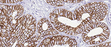
Antibody staining in 20 different cancers is summarized by a selection of four standard cancer tissue samples representative of the overall staining pattern. From left: colorectal cancer, breast cancer, prostate cancer and lung cancer. An additional fifth image can be added as a complement. The assay and annotation is described here. Note that samples used for immunohistochemistry by the Human Protein Atlas do not correspond to samples in the TCGA dataset. For histological reference, visit the histological dictionary.
For each cancer, color-coded bars indicate the percentage of patients (maximum 12 patients) with high and medium protein expression level. The cancer types are color-coded according to which type of normal organ the cancer originates from. Low or not detected protein expression results in a white bar. Mouse-over function shows details about expression level and normal tissue of origin. The images and annotations can be accessed by clicking on the cancer name or protein expression bar. If more than one antibody is analyzed, the tabs at the top of the staining summary section can be used to toggle between the different antibodies.
Organ
Expression
Alphabetical
HPA018420
Several cancers displayed moderate to strong cytoplasmic and nuclear staining in a fraction of cells, including cervical and endometrial cancers. Testis, colorectal and thyroid cancers and gliomas were negative.
We use cookies to enhance the usability of our website. If you continue, we'll assume that you are happy to receive all cookies. More information. Don't show this again.


 The Human Protein Atlas project is funded