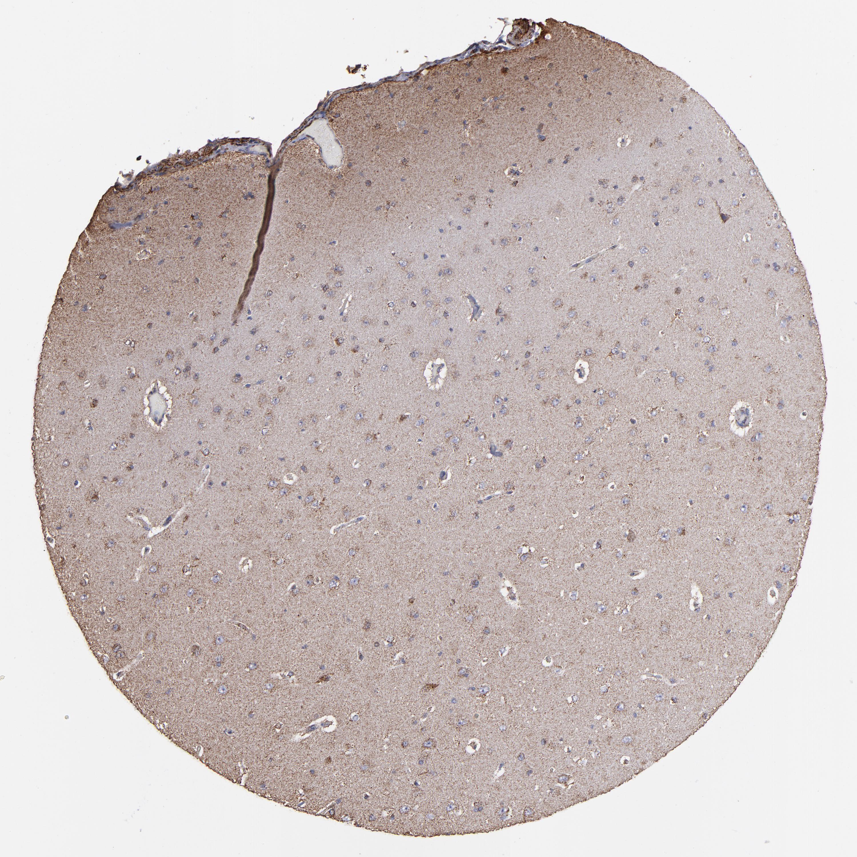
On the top, protein expression in current human tissue, based on all annotated cell types, is reported with the units not detected (n), low (l), medium (m) and high (h). Underneath, protein expression in each annotated cell type are reported using the same units. Protein expression data is based on knowledge-based annotation. For genes where more than one antibody has been used, a collective score is set. If knowledge-based annotation could not be performed for a gene, no data is displayed here. View antibody staining data further down this page.
h
m
l
n
Endothelial cells:
Medium
Glial cells:
Medium
Neuronal cells:
High
Neuropil:
Medium
RNA expressioni
A summary of mRNA expression data available for current tissue based on several datasets. The mRNA expression levels in human tissues are based on RNA-seq data generated by the Human Protein Atlas HPA, Genotype-Tissue Expression GTEx portal and CAGE data generated by the FANTOM5 consortium. Consensus normalized expression levels for human tissues was created by combining the data from HPA and GTEx datasets. The mRNA expression levels in pig brain are based on RNA-seq data generated by the Human Protein Atlas, and for mouse brain HPA data and in situ hybridization generated by the Allen brain atlas dataset are reported.
Scroll down to view mRNA expression data in more detail.
Consensus:
53.1
nTPM
HPA:
53.1
nTPM
HPA Brain:
378.6
nTPM
HPA PFC Brain:
18.0
nTPM
FANTOM5:
318.0
Scaled Tags Per Million
Macaque:
113.8
nTPM
Mouse:
55.7
nTPM
Rat:
72.2
nTPM
Pig:
85.6
nTPM
CEREBRAL CORTEX - Antibody stainingi
Antibody staining in the annotated cell types in the current human tissue is reported as not detected, low, medium, or high, based on conventional immunohistochemistry profiling in selected tissues. This score is based on the combination of the staining intensity and fraction of stained cells. Each image is clickable and will lead to virtual microscopy that enables deeper exploration of all samples and also displays staining intensity scores, fraction scores and subcellular localization as well as patient and tissue information for each sample.
Ascending and descending describe two types of gradient, based on difference in staining intensity between enterocytes in crypts and apical enterocytes.
Ascending - the staining intensity is stronger in apical enterocytes (towards the lumen) compared to enterocytes in the crypts (gland based)
Descending - the staining is weaker in apical enterocytes compared to enterocytes in crypts
Antibody HPA001814
Endothelial cells
Medium
Glial cells
Medium
Neuronal cells
High
Neuropil
Medium
CEREBRAL CORTEX - PROTEINTALKS DB MS
Median intensity
66104
Sample
Intensity
Cerebral cortex_01
73217
Cerebral cortex_02
66691
Cerebral cortex_03
65517
Cerebral cortex_04
57754
Cerebral cortex_05
56847
Cerebral cortex_06
76894
Cerebral cortex_07
54742
Cerebral cortex_08
61566
Cerebral cortex_09
47704
Cerebral cortex_10
81605
Cerebral cortex_11
40498
Cerebral cortex_12
89657
Cerebral cortex_13
60955
Cerebral cortex_14
43721
Cerebral cortex_15
110471
Cerebral cortex_16
63762
Cerebral cortex_17
41343
Cerebral cortex_18
48342
Cerebral cortex_19
98698
Cerebral cortex_20
119595
Cerebral cortex_21
52370
Cerebral cortex_22
61046
Cerebral cortex_23
35766
Cerebral cortex_24
119717
Cerebral cortex_25
119719
Cerebral cortex_26
111632
Cerebral cortex_27
106338
Cerebral cortex_28
117547
Cerebral cortex_29
80947
Cerebral cortex_30
87062
Cerebral cortex_31
77790
Cerebral cortex_32
90429
Cerebral cortex_33
48474
Cerebral cortex_34
75798
Cerebral cortex_35
69330
Cerebral cortex_36
54505
Cerebral cortex_37
56688
Cerebral cortex_38
63562
CEREBRAL CORTEX - MANN MS
Median intensity
209244604
Replicate
Intensity
Cerebral cortex_01
209244604
Cerebral cortex_02
195304258
Cerebral cortex_03
266212237
CEREBRAL CORTEX - HPA RNA-seqi
The RNA-seq details section shows detailed information about the individual samples used for the transcript profiling and results of the RNA-seq analysis.
Information about each individual sample is listed below, including gender, age, a tissue section image and estimated fractions of cell types. nTPM (normalized transcripts per million) values give a quantification of the gene abundance which is comparable between different genes and samples.
Average nTPMi
Max subtype nTPM is the value for the subtype with the largest average nTPM across the subtype samples.
53.1
Male, age 5
Cerebral cortex sample 48 nTPM: 41.4
Female, age 40
Cerebral cortex sample 105 nTPM: 24.8
Cell types%
Neuronal cells:
35
Glial cells and endothelial cells:
65
Male, age 70
Cerebral cortex sample 106 nTPM: 93
Cell types%
Neuronal cells:
35
Glial cells and endothelial cells:
65
CEREBRAL CORTEX - HPA Brain Stereo-seq
CEREBRAL CORTEX - HPA Brain RNA-seq
Max subtype nTPMi
Max subtype nTPM is the value for the subtype with the largest average nTPM across the subtype samples.
RNA expression in human tissues obtained through Cap Analysis of Gene Expression (CAGE) generated by the FANTOM5 project are reported as Scaled Tags Per Million.
Max subtype Scaled Tags Per Millioni
Max subtype Scaled Tags Per Million is the value for the subtype with the largest average Scaled Tags Per Million across the subtype samples.
Max subtype nTPM is the value for the subtype with the largest average nTPM across the subtype samples.
113.8
Macaque sample
Sample description
nTPM
Angular gyrus nTPM: 113.3
Samples: 1
Max nTPM: 113.3
Min nTPM: 113.3
Sample A
113.3
Claustrum nTPM: 83.8
Samples: 1
Max nTPM: 83.8
Min nTPM: 83.8
Sample B
83.8
Fusiform gyrus nTPM: 86.8
Samples: 2
Max nTPM: 94.4
Min nTPM: 79.1
Sample A
79.1
Sample B
94.4
Inferior temporal gyrus nTPM: 81.2
Samples: 2
Max nTPM: 93.2
Min nTPM: 69.1
Sample A
69.1
Sample B
93.2
Middle temporal gyrus nTPM: 101.3
Samples: 2
Max nTPM: 108.7
Min nTPM: 93.9
Sample A
108.7
Sample B
93.9
Postcentral gyrus nTPM: 106.1
Samples: 2
Max nTPM: 123.3
Min nTPM: 88.9
Sample A
88.9
Sample B
123.3
Posterior cingulate cortex nTPM: 95.0
Samples: 2
Max nTPM: 96.9
Min nTPM: 93.0
Sample A
93.0
Sample B
96.9
Precentral gyrus nTPM: 113.8
Samples: 3
Max nTPM: 124.0
Min nTPM: 107.4
Sample A
110.0
Sample B
124.0
Sample C
107.4
Superior parietal lobule nTPM: 100.8
Samples: 2
Max nTPM: 103.8
Min nTPM: 97.8
Sample A
97.8
Sample B
103.8
Superior temporal gyrus nTPM: 85.0
Samples: 2
Max nTPM: 87.0
Min nTPM: 83.0
Sample A
83.0
Sample B
87.0
Supramarginal gyrus nTPM: 105.4
Samples: 2
Max nTPM: 106.0
Min nTPM: 104.8
Sample A
106.0
Sample B
104.8
Gyrus rectus nTPM: 85.9
Samples: 2
Max nTPM: 88.6
Min nTPM: 83.2
Sample A
83.2
Sample B
88.6
Insular cortex nTPM: 82.9
Samples: 2
Max nTPM: 85.9
Min nTPM: 79.8
Sample A
85.9
Sample B
79.8
Superior frontal gyrus nTPM: 108.4
Samples: 2
Max nTPM: 119.1
Min nTPM: 97.6
Sample A
97.6
Sample B
119.1
CEREBRAL CORTEX - Pig RNA-seqi
RNA-seq data obtained from pig brain is reported as nTPM. Normal distribution across the dataset is visualized with box plots, shown as median and 25th and 75th percentiles. Points are displayed as outliers if they are above or below 1.5 times the interquartile range. nTPM values of the individual samples are presented next to the box plot.
Max subtype nTPMi
Max subtype nTPM is the value for the subtype with the largest average nTPM across the subtype samples.
85.6
Pig sample
Sample description
nTPM
Occipital cortex nTPM: 77.9
Samples: 4
Max nTPM: 86.9
Min nTPM: 68.1
Sample A
Female
86.9
Sample B
Female
68.1
Sample C
Male
85.6
Sample D
Male
71.0
Retrosplenial cortex nTPM: 68.3
Samples: 4
Max nTPM: 78.9
Min nTPM: 62.2
Sample A
Female
69.4
Sample B
Female
62.2
Sample C
Male
78.9
Sample D
Male
62.8
Cingulate cortex nTPM: 71.6
Samples: 4
Max nTPM: 81.5
Min nTPM: 58.6
Sample A
Female
81.5
Sample B
Female
58.6
Sample C
Male
78.2
Sample D
Male
67.9
Insular cortex nTPM: 60.1
Samples: 4
Max nTPM: 74.9
Min nTPM: 49.6
Sample A
Female
74.9
Sample B
Female
49.6
Sample C
Male
62.2
Sample D
Male
53.8
Motor cortex nTPM: 80.2
Samples: 4
Max nTPM: 91.7
Min nTPM: 68.5
Sample A
Female
89.3
Sample B
Female
68.5
Sample C
Male
91.7
Sample D
Male
71.1
Prefrontal cortex nTPM: 71.6
Samples: 4
Max nTPM: 76.3
Min nTPM: 68.7
Sample A
Female
76.3
Sample B
Female
70.2
Sample C
Male
71.0
Sample D
Male
68.7
Somatosensory cortex nTPM: 85.6
Samples: 3
Max nTPM: 97.7
Min nTPM: 73.7
Sample B
Female
73.7
Sample C
Male
85.3
Sample D
Male
97.7
Temporal cortex nTPM: 68.5
Samples: 3
Max nTPM: 72.2
Min nTPM: 63.2
Sample A
Female
72.2
Sample B
Female
63.2
Sample D
Male
70.1
CEREBRAL CORTEX - Mouse RNA-seqi
RNA-seq data obtained from mouse brain is reported as nTPM. Normal distribution across the dataset is visualized with box plots, shown as median and 25th and 75th percentiles. Points are displayed as outliers if they are above or below 1.5 times the interquartile range. nTPM values of the individual samples are presented next to the box plot.
Max subtype nTPMi
Max subtype nTPM is the value for the subtype with the largest average nTPM across the subtype samples.
55.7
Mouse sample
Sample description
nTPM
Motor and prefrontal cortex nTPM: 45.7
Samples: 4
Max nTPM: 49.0
Min nTPM: 42.0
Sample A
49.0
Sample B
45.4
Sample C
46.5
Sample D
42.0
Retrosplenial and cingulate cortex nTPM: 55.7
Samples: 3
Max nTPM: 59.8
Min nTPM: 49.6
Sample A
49.6
Sample B
59.8
Sample C
57.7
Somatosensory cortex nTPM: 40.7
Samples: 4
Max nTPM: 44.9
Min nTPM: 37.6
Sample A
44.9
Sample B
38.6
Sample C
41.8
Sample D
37.6
Visual cortex nTPM: 49.5
Samples: 4
Max nTPM: 55.5
Min nTPM: 46.5
Sample A
48.2
Sample B
46.5
Sample C
55.5
Sample D
47.8
CEREBRAL CORTEX - Rat RNA-seq
Max subtype nTPMi
Max subtype nTPM is the value for the subtype with the largest average nTPM across the subtype samples.
72.2
Rat sample
Sample description
nTPM
Occipital cortex nTPM: 70.2
Samples: 4
Max nTPM: 77.7
Min nTPM: 65.5
Sample A
65.7
Sample A
65.5
Sample A
77.7
Sample A
71.9
Retrosplenial and cingulate cortex nTPM: 72.2
Samples: 4
Max nTPM: 73.4
Min nTPM: 70.8
Sample A
73.4
Sample A
73.4
Sample B
70.8
Sample A
71.1
Somatosensory cortex nTPM: 72.1
Samples: 4
Max nTPM: 76.0
Min nTPM: 69.4
Sample A
70.3
Sample A
76.0
Sample A
72.7
Sample A
69.4
We use cookies to enhance the usability of our website. If you continue, we'll assume that you are happy to receive all cookies. More information. Don't show this again.